like hip dysplasia, canine elbow dysplasia is more common in large breeds. What are its symptoms and treatment?
and
mainly affect large dogs. Elbow dysplasia is usually characterized by obvious claudication from childhood. Some breeds are prone to this abnormality, partly due to heredity.
and
elbow joints are composed of three bones: humerus, radius and ulna. These bones have some processes, and their abnormalities define dysplasia, so it is important to find them on X-rays. Elbow dysplasia in dogs is a developmental abnormality. With the increase of growth, it can lead to pain and posture disorders, including claudication. These abnormalities lead to joint disharmony, followed by elbow osteoarthritis.
appear in puppies, mainly large dogs. In addition, the incidence of males is twice that of females.
Some large breeds do tend to have elbow dysplasia, which is bilateral in most cases. These include the Rottweiler, Berne shepherd, Labrador Retriever, golden retriever, Saint Bernard, German Shepherd and mastiff, Staffordshire Terrier, dog and basset. Elbow dysplasia is sometimes associated with femoral hip dysplasia.
The etiology of
elbow dysplasia involves multiple genes, and canine elbow dysplasia has some genetic causes. However, dogs carrying these genes may not see this abnormal development. Dislocation of the surface of radius and ulna leads to too short radius or ulna, abnormal friction between radius and ulna, abnormal friction between humerus and ulna
and other factors may also lead to the deterioration of elbow dysplasia, Due to excessive and repeated exercise and physical labor, inappropriate diet, including excessive energy intake and accelerated growth.
therefore, it is important to ensure that puppies are fed in a balanced manner, There is no excessive or lack of physical activity. There are four forms of elbow dysplasia in
. Elbow dysplasia in
dogs can be divided into four types:
Osteochondritis exfoliatum: this form of elbow dysplasia is characterized by joint inflammation and pain caused by the separation of a piece of necrotic cartilage from the medial side of the protruding joint surface of the humerus. Anal process nonunion: during the growth of healthy puppies, a bone at the back of the elbow must fuse with the ulna (one of the two bones of the forearm radius) around the 16th week. When the elbow is subjected to abnormal stress, the combination of this process does not occur, and then NUPA type elbow dysplasia (non combination of elbow process): non combination leads to the conflict between humerus and ulna, and the elbow process is stuck, Fragmentation of the coronal process: synovitis (arthritis) is a small piece of bone fragment separated from the inner surface of the ulnar joint. This may lead to joint surface irritation and humeral cartilage erosion.Uncoordinated elbow joint: when the joint surface embolization fails or is incomplete, it will lead to osteoarthritis. This form of elbow dysplasia in dogs is called elbow disharmony. Disharmony can be radioulna, humeral ulna or radioulna. The four lesions of
may sometimes be related to each other.
symptoms and treatment of
Elbow dysplasia in dogs is usually characterized by chest limb claudication. Lameness may occur as early as 5 months in exercise, or it may be a simple exercise. Lameness may vary from individual to individual: it may be sudden, it may be latent, it may be intermittent, or it may be permanent. However, dysplasia may also be detected only in adulthood.
in some puppies, postural deviation and running cuts can be observed to reduce pain.
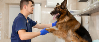
in order to determine the diagnosis, veterinarians conducted postural studies on animals, Orthopaedic examination and X-ray examination. It can also scan the elbow. When touching the limbs, the veterinarian will notice the depression of the elbow, and the bending will be reduced when moving. The Campbell test conducted by the veterinarian was for the pain of elbow dysplasia.
treatment is mainly surgical operation, and its effectiveness depends on the degree of abnormality and early diagnosis and treatment.
for young people, the operation is carried out under arthroscopy, Cartilage fragments should be removed to stimulate bone healing. If the animal is an adult with severe osteoarthritis, the treatment is not surgical, but medical, using non steroidal anti-inflammatory drugs and cartilage protectors. Animals must lose weight and take appropriate physical exercise to reduce the pressure on their joints. If the animal does not respond to drug treatment, intra-articular injection or electric shock can be performed. In France, there are very professional surgical treatments, but they are still rarely available.
and
are performed under anesthesia after operation, especially to reduce pain. Postoperative claudication did not disappear immediately; Only 30% of dogs will never limp again, but 85% of dogs will improve.
and
require screening dogs belonging to dangerous breeds, because the treatment effect will be better if they are treated early. “Kdspe


