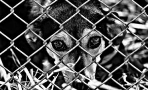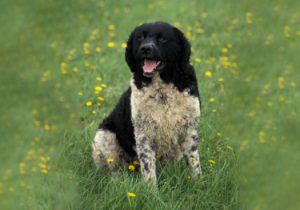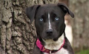
histiocytosis is a group of different diseases that affect almost all dog breeds. Skin histiocytoma, reactive histiocytoma and malignant histiocytoma.
in addition to skin histiocytoma, which is very common in dogs, other forms of histiocytoma are quite rare.
histiocytoma is an immune cell located in connective tissue, Macrophages will first contact pathogens. These cells present antigens (part of pathogens) to lymphocytes to activate them and protect the body from attack.
malignant histiocytosis
malignant histiocytosis, also known as histiocytic sarcoma (SH) (local or disseminated), It is a very rare cancer.
localized histiocytic sarcoma is a mass, usually close to the joint, related to tissue cells in connective tissue.
mainly affects Berne shepherds, golden retrievers, Labradors, flat coated hounds and Rottweilers aged 2 to 15.
This form of histiocytosis does not affect the skin, but affects different internal organs (kidney, liver, spleen, lung, etc.). Berne shepherd dogs are highly susceptible to this type of tumor, and there is a special genetic test (HS test), which is sold to breeders by major laboratories. The local HS of
and
can be identified by the presence of a mass on the skin surface, Then transfer will lead to other general clinical symptoms, such as high fever (fever), cough and dyspnea, indifference (extreme fatigue), anorexia, so as to lose weight. Some people may also have mobility difficulties. In decentralized HS, these clinical symptoms will lead the owner to consult the veterinarian, mainly internally.
can be diagnosed by imaging (ultrasound, MRI) or tissue sampling for histological analysis (tissue analysis).
Unfortunately, there is no cure; Veterinarians can only extend your dog’s life through chemotherapy to ensure that they have a good quality of life for the rest of the day.
reactive histiocytosis
reactive histiocytosis has two different forms: skin and system.
skin tissue cell proliferation is characterized by skin plaques up to 4 cm, sometimes without hair. Please note that the injury does not cause pain or itching. This is a non hereditary and rare histiocytosis, especially affecting the
golden retriever, German Shepherd and shepherd
. The prognosis of this disease is usually favorable. Nearly 50% of dogs responded to treatment.
and
systemic histiocytes proliferated irregularly and developed for a long time. This disease mainly affects Bernie collies and to a lesser extent
Labrador, Rottweiler, Doberman, Belgian shepherd, Iraqi otter, poodle, border collie, golden retriever Cross
such as skin tissue cell proliferation, Systemic histiocytosis can also affect the skin, but it can also affect other organs, such as the nose, eyes, lungs, liver, bone marrow, spleen and kidney. It is usually accompanied by:Kdspe “weight loss, anorexia and sleepiness”
unfortunately, this disease has a poor response to treatment due to its chronic form. The average survival time of dogs is 9 to 10 months (Dobson and Duncan 2003 – Ettinger 2000 – Paterson et al., 1995 – Vail 2001). “Difficult diagnosis and rare diseases are rarely the primary assumptions of differential diagnosis. Therefore, later studies were carried out through biopsy and histological analysis, ultrasound and MRI… In view of the poor response to treatment, this has no special impact on the prognosis.
skin histiocytoma
skin histiocytoma is a kind of skin tumor, Usually benign. It forms a painless round nodule between 1 and 2 cm in diameter. However, it may ulcerate because its surface is quite fragile. It especially affects puppies (under 3 years old). Therefore, this feeling decreases with age. The most seriously affected area in animals is
facial extremities end ear Pavilion
. The most vulnerable species are:
Boxer, grapefruit, German dog, rooster, Shetland shepherd, bull terrier, shnauzer, epagneuls
Diagnosis should be made by cell function (puncture of the mass with a needle) and cytology (analysis of the cells that make up the mass). Sometimes biopsy is required for histological analysis, which is carried out by a professional laboratory. The lesions of cutaneous histiocytoma usually disappear spontaneously. Some forms, more or less complex and persistent, require antibiotics or surgery. The prognosis of this condition is very good.